BLACK FRIDAY IS ON! UP TO 25% OFF + FREE 7-PIECE GIFT WITH $75+

SHOP NOW

Subscribe to our newsletter

Get 10% off your first order, plus the latest updates and news!

Normal, dry to atopic skin

Eczema on black skin explained: causes, symptoms, and care

Reading time : 8 min

dark skin woman scratching

Eczema on black skin explained: causes, symptoms, and care


Key takeaways from this guide


  • Eczema on black skin presents differently from lighter complexions, with the classic redness replaced by grey, purple or brown tones, so early diagnosis requires careful clinical attention.
  • Atopic dermatitis affects Black populations in the United States more frequently and tends to be more severe, and standard scoring tools that rely on visible erythema underestimate the real picture.
  • Post-inflammatory hyperpigmentation is the most common lasting complication, and dark spots can persist for months after the flare itself has cleared.
  • Daily moisturizing with fragrance-free, lipid-rich emollients is the basis of care, while prescription anti-inflammatory treatments handle more intense cases.
  • Sun protection limits the pigmentation marks that follow flares, and gentle skincare helps prevent new ones.


Eczema on black skin is often missed or misdiagnosed for a simple reason. Clinicians are trained to look for redness, and that redness shows up very differently on darker complexions.
 

Inflammation reads as bright red on light skin. On darker tones, the same inflammation can appear grey, purple, brown or even ashen. Severity scoring tools designed decades ago on predominantly light-skinned populations underestimate what is actually happening at the skin level.
 

This guide covers how eczema actually presents on black skin, explains why standard assessment often falls short, and describes the care strategies that work. The information is based on peer-reviewed dermatological literature focused on skin of color.


Introduction to eczema on black skin


How eczema appears on darker complexions


Eczema is an inflammatory skin condition, and the inflammation itself is identical across skin tones. What changes is how it manifests visibly at the surface.

On lighter complexions, eczema typically appears as pink or red patches. On black skin, the same inflammation shows differently, in shades of grey and sometimes purple, brown or ashen tones. The visual contrast that practitioners are taught to identify is muted or absent.

Clinical presentations also differ in texture. Lesions appear more papular and lichenified; follicular accentuation is common, and dermatologists familiar with skin of color recognize these features as core diagnostic signs in this population1.


Prevalence and severity in Black populations


Atopic dermatitis is not equally distributed across populations. Prevalence is higher among Black children in the United States than among white children, and severity tends to be greater as well1. In a nationally representative survey of 102,353 US children, 10.7% had eczema, and Black children were significantly more likely to be affected (odds ratio 1.70)6.
 

Several factors contribute, including genetic predisposition and differences in skin barrier composition. Diagnosis is often delayed when visible signs do not match textbook descriptions, and access to dermatologists trained on skin of color is also limited in the United States healthcare system.


Common symptoms and signs on black skin



Itching is the same on every skin tone, intense and disruptive, and frequently more pronounced at night.
 

What differs is what surrounds the itch. On black skin, the inflamed area looks darker than the surrounding skin rather than redder, and it may feel warm without visibly flushing. Many patients scratch for weeks before the condition is correctly identified.
 

Repeated scratching thickens the skin, a process of dermatologists calls lichenification, and on darker complexions the thickening can deepen to nearly black in chronic patches.
 

Two pigment changes typically follow eczema flares on black skin. Hyperpigmentation leaves dark spots after the inflammation has cleared, while hypopigmentation produces lighter patches when pigment cells are more deeply affected.
 

Post-inflammatory hyperpigmentation is the most frequent and most lasting complication in patients with skin of color2, and the marks can persist for months after the underlying eczema clears.
 

Severity scoring tools were developed on light skin. EASI, SCORAD and IGA all rely heavily on visible erythema, which is muted on black skin, so the score comes out lower than the clinical reality.
 

Specific recommendations exist for managing atopic dermatitis in patients with skin of color3, and a growing body of dermatological literature now advocates for revised assessment tools. Until those become standard practice, patients and parents benefit from advocating based on symptoms rather than visual scoring alone.
 

Common causes and triggers


Most eczema on black skin is atopic dermatitis. The underlying mechanism is identical, and only the visible presentation differs.
 

Skin barrier dysfunction sits at the core of the condition. Filaggrin gene mutations are documented across populations; ceramide profiles differ between ethnic groups, and immune response variations also contribute1. The combined effect is faster water loss through the skin, irritants penetrate more easily, and the immune system overreacts to common triggers.
 

Common triggers include stress, dry air, and exposure to irritants. Identifying personal triggers requires consistent observation over time. For broader context on the condition itself, see eczema.
 

Contact dermatitis is the second main cause of eczema-like presentations on black skin.

Hair products are a frequent culprit in this population, particularly relaxers and dyes, and fragranced oils applied close to the scalp can affect the face and neck. Nickel jewelry, fragranced laundry detergents, and certain cosmetics also trigger reactions. The reaction mechanisms are identical to those seen on other skin tones, but the visible signs are not.
 

Patch testing identifies the specific allergens behind contact reactions when this etiology is suspected.
 

Effective treatment options for black skin


Daily moisturizing matters more on skin prone to dryness and ashy texture. Twice-daily application of emollients reduces flare frequency and limits the need for stronger treatments. The benefit of consistent emollient use is well established4.
 

Ointments and rich creams work better than lighter lotions on textured skin and during winter months. The application window matters as much as the product itself, since absorption is optimal within two minutes of bathing, while the skin is still slightly damp.
 

Topical corticosteroids are the first-line prescription option. Calcineurin inhibitors such as tacrolimus or pimecrolimus serve as alternatives, useful on the face and skin folds. Their relative efficacy has been reviewed in a recent Cochrane network meta-analysis of 291 trials and 45,846 participants5.
 

On areas prone to hyperpigmentation, starting treatment early protects pigment integrity, whereas waiting for spontaneous resolution adds weeks of melanin imbalance. The principle is direct, as shorter flares produce less pigmentation.
 

Once dark spots have formed, they take time to fade, and there is no shortcut.
 

Topical options include azelaic acid and niacinamide, vitamin C contributes to brightening, and retinoids work in specific cases under dermatology supervision. None produce overnight results, and most require 8 to 12 weeks for visible improvement. Consistent sun protection during this period prevents the marks from deepening.
 

Stronger options exist under dermatology prescription, but preventing the next flare matters more than treating the marks from the last one.
 

Preventive measures and lifestyle adjustments


A consistent routine produces better results than any single product. It combines short lukewarm baths, mild fragrance-free cleansers, patting dry rather than rubbing, and moisturizer applied within two minutes.
 

Adjustments come from observing what triggers each individual. Hair products that touch the face, laundry detergents used on bedding and clothing, and even the cleansing products used by other family members in shared bathrooms can all have an impact.
 

Black skin produces more melanin than lighter complexions, but UV-induced pigmentation still happens, and it is often more visible.
 

Daily broad-spectrum SPF 30 limits the worsening of post-inflammatory hyperpigmentation. Mineral sunscreens with zinc oxide or titanium dioxide are well tolerated, though they can leave a white cast that tinted mineral formulations and modern non-nano versions resolve. Reapplication every two hours during outdoor exposure matters more than brand choice.
 

Black skin produces more melanin than lighter complexions, but UV-induced pigmentation still happens, and it is often more visible.
 

Daily broad-spectrum SPF 30 limits the worsening of post-inflammatory hyperpigmentation. Mineral sunscreens with zinc oxide or titanium dioxide are well tolerated, though they can leave a white cast that tinted mineral formulations and modern non-nano versions resolve. Reapplication every two hours during outdoor exposure matters more than brand choice.
 

Long-term management strategies


Not all dermatologists have equivalent training on skin of color, and the gap shows up in both diagnostic accuracy and treatment selection.
 

Online directories sometimes filter for this expertise, and recommendations from Black dermatology professional organizations also help. When previous consultations underestimated the symptoms based on visible signs alone, a practitioner experienced with darker complexions often changes the picture.
 

Not all dermatologists have equivalent training on skin of color, and the gap shows up in both diagnostic accuracy and treatment selection.
 

Online directories sometimes filter for this expertise, and recommendations from Black dermatology professional organizations also help. When previous consultations underestimated the symptoms based on visible signs alone, a practitioner experienced with darker complexions often changes the picture.
 

Most eczema cases respond to consistent topical care, but some do not.
 

Persistent flaring despite daily moisturizing and prescribed treatment indicates that the current approach needs adjustment. Sleep deeply affected by itching, secondary bacterial infections, and notable emotional impact are all reasons to revisit the treatment plan. Newer systemic treatments now exist for more intense atopic dermatitis that responds less to topical therapy alone.
 

woman with atoderm intensive balm packshot

ATODERM skincare for atopic-prone skin of color


On darker skin tones, flares can leave lasting pigmentation, so calming inflammation quickly and keeping the barrier hydrated every day are central to limiting marks. Fragrance-free, lipid-rich emollients suit the dryness and ashy texture that often accompany eczema-prone skin of color.


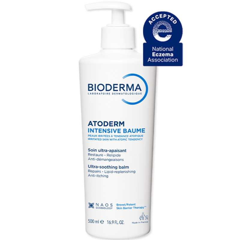
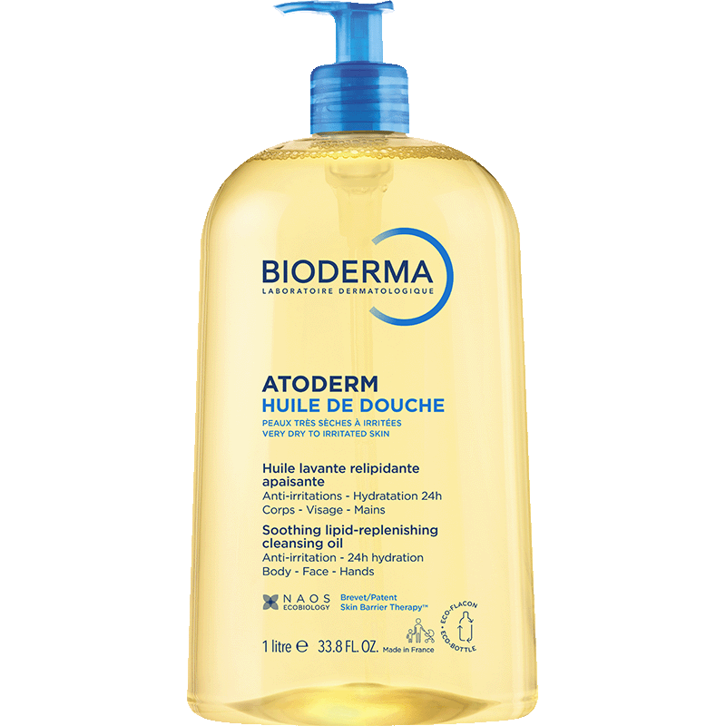
Within the ATODERM range, Atoderm Intensive Balm is specially formulated for very dry to atopic-prone skin, helping provide long-lasting hydration and skin comfort. For daily cleansing, Atoderm Shower Oil gently cleanses without stripping the skin of essential moisture, helping maintain hydration and leaving skin feeling soft and nourished. Both products are fragrance-free.

Common questions about eczema on black skin


Yes. Post-inflammatory hyperpigmentation is the most common long-term consequence of eczema flares on darker skin. The dark spots result from inflammation interfering with normal melanin production and can persist for months after the underlying eczema has resolved.
 

Treating eczema flares early is the most effective way to limit them.
 

Atopic dermatitis prevalence and intensity are both higher in Black populations in the United States. Some of this is biological, since genetic differences and barrier function variations are documented across ethnic groups. Other factors are access-related, as diagnosis is often delayed because the clinical signs present differently and require more clinical experience to identify on darker complexions.
 

Skin with higher melanin content does not display erythema in the same way. The inflammation is real, but it presents through grey or purple shading, sometimes brown or violaceous, rather than the typical pink or red. Standard severity scoring tools based on visible redness systematically underestimate eczema on black skin.
 

This is why specialized dermatological assessment matters in this population.
 

Scientific references

  1. Kaufman BP, Guttman-Yassky E, Alexis AF. Atopic dermatitis in diverse racial and ethnic groups - Variations in epidemiology, genetics, clinical presentation and treatment. Exp Dermatol. 2018;27(4):340-357. https://pubmed.ncbi.nlm.nih.gov/29457272/
  2. Kaufman BP, Aman T, Alexis AF. Postinflammatory Hyperpigmentation: Epidemiology, Clinical Presentation, Pathogenesis and Treatment. Am J Clin Dermatol. 2018;19(4):489-503. https://pubmed.ncbi.nlm.nih.gov/29222629/
  3. Lebovits R, Lara-Corrales I, Pope E. Dermatology: how to manage atopic dermatitis in patients with skin of colour. Paediatr Child Health. 2022;27(6):371-373. https://pubmed.ncbi.nlm.nih.gov/35720054/
  4. Van Zuuren EJ, Fedorowicz Z, Christensen R, Lavrijsen APM, Arents BWM. Emollients and moisturisers for eczema. Cochrane Database Syst Rev. 2017;2(2):CD012119. https://pubmed.ncbi.nlm.nih.gov/28166390/
  5. Lax SJ, Van Vogt E, Candy B, et al. Topical anti-inflammatory treatments for eczema: network meta-analysis. Cochrane Database Syst Rev. 2024;8(8):CD015064. https://pubmed.ncbi.nlm.nih.gov/39105474/
  6. Shaw TE, Currie GP, Koudelka CW, Simpson EL. Eczema prevalence in the United States: data from the 2003 National Survey of Children's Health. J Invest Dermatol. 2011;131(1):67-73. https://pubmed.ncbi.nlm.nih.gov/20739951/


Author

Content written by the BIODERMA USA editorial team and medically reviewed by Anaïs Caldirola, Medical lead at NAOS, ensuring accuracy and scientific reliability. Medical information is based on established dermatological knowledge and peer-reviewed sources.